
09 Jul Heel Pain
Heel Pain Series Week 1: Anatomy
https://www.sportspodiatry.com.au/services/
Heel pain is one of the most common conditions we treat as Podiatrists. Most people describe pain on the bottom heel bone (the calcaneus) on the medial side (the side closest to the midline).
I have often heard people describe the pain as a bone bruise/stone bruise or really deep pain. It hurts so bad when they get out of bed in the morning or after they have been sitting for a while, and they hobble along until it warms up a little bit.
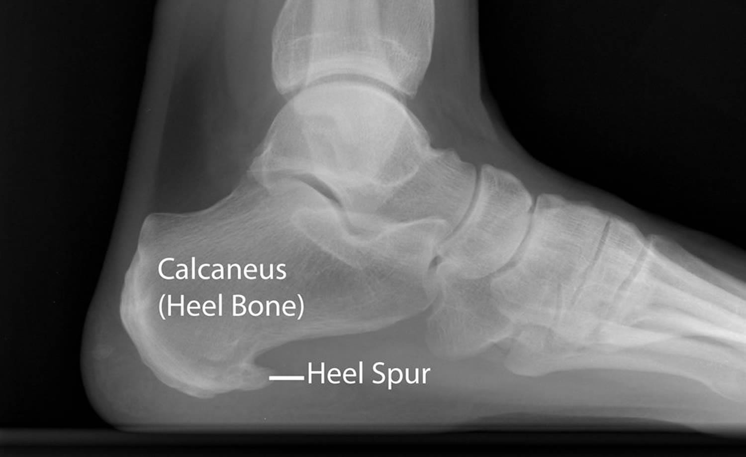
The first thing people (and their doctors) seem to think they have is a heel spur. So let us look deeper at the bones for a moment. Sometimes, but not always, an X-Ray report will confirm a “spur” at the medial tubercle of the calcaneus. You can see an example of this in the picture above that I have borrowed (with permission) from one of my patients.
My patients tell me they are surprised the “spur” is not vertical because it feels like it is stabbing right down! Would you believe this dagger-like bit of bone may have nothing to do with your pain? The patient above actually has no pain in her heel at all! More on this later.
Above the heel bone sits the talus, and the cuboid in front of it. But there is more to a heel than just the bones we can see on X-Ray.
What else is in this area?
If you peel back the skin, the next layer you find is the lesser-known facia.
Every. Single. Day. I have someone come into my office telling me they have plantar fasciitis.
The Plantar fascia is an aponeurosis. “A whaaatttt?!” you say. It is not a tendon, it is not a muscle, it is not a ligament, it’s something else entirely! It is made up of type 1 collagen, a few elastin fibres, reticulin, and ground substance (mucous that glues the facia together). It is avascular (not very good blood supply) and the part on the foot is not super-elastic.
Another name for it is sinew. For you meat eaters out there, you will know what I mean. It is the chewy, tough stuff that you don’t eat, the white stuff you see when you are taking the skin off a chicken breast. Its job is to hold in the little muscles of the bottom side of the foot and helps structurally hold up that arch. It attaches from the inside of the heel bone (calcaneus), and you can break it down into 3 sections.
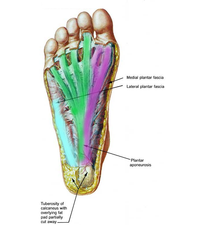
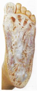
Photo credit: https://profacas.wordpress.com/2014/05/ with modifications by Hannah Salsbury on the left. On the right is a human cadaver (what it looks like!) from McMinn, M.H, Hutchings, R. T, Logan, B.M, Colour Atlas of Foot and Ankle Anatomy 2nd edition, Mosby-Wolfe, Spain.
There is a central band (green), a lateral band (light blue), and a medial band (purple). The plantar fascia runs to the toes and inserts deep inside the foot. Superficially it inserts into your tootsies and the deepest layer of skin of the ball of the foot where all the sensory receptors are (making it sensitive). The plantar fascia acts kind of like an anchor to the skin and supports it from shearing forces. It allows everything to slide and glide over each other…except when it is stressed or damaged!
Facia surrounds and separates every musculoskeletal structure in the body and it’s all interlinked like a big web, so it’s hard to look at the plantar part all on its own, it’s part of a bigger interconnected system.
The Fat Pad:
The cushion under your heel bone is made of fat. It absorbs the shock and stress of ground reaction forces, particularly during heel strike (heel contact during walking). The force and impact that your feet absorb when you walk is 2.5 times the weight of your body. When you run or jump, that impact increases even more!
We know as we get older this fatty tissue tends to get less and less cushiony and elastic. The fat pad of the heel can atrophy (shrink), become worn and thin, or become inflamed and painful, resulting in heel fat pad syndrome.
The tiny (intrinsic) muscles of the foot:
There are muscles, little tiny muscles in the foot and bigger stronger muscles that attach outside the foot. The little ones you have probably never thought about before. They are more important than you might think.
Your abductor hallucis, flexor digitorum brevis and quadratus plantae are part of your balance system. They have been measured to work harder with increased postural demand (Kelly et al. 2011). Remember when you went stand-up paddle boarding for the first time? Are feet sore after walking on the soft sand at the beach? All you have to do is try standing on one leg for a while, you will feel these little guys working hard!
They also help our foot shock absorb when the foot hits the ground, and they turn the foot into a spring during toe-off. Look where they attach…yep…back at the heel and they flow right through that arch area. Now, these I am pointing out are just the biggest of the intrinsic muscles, there are 18 of them in total in the foot!
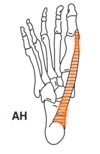
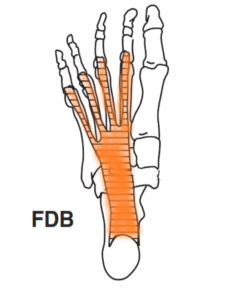
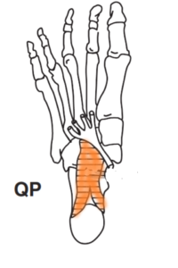
Pictures credit: L. Kelly, S, Kuitunen, Racinais, A, Cresswell. Recruitment of Plantar Intrinsic Foot Muscles with Increasing Postural Demand. Clinical Biomechanics 27 (2012) 46-51
The bigger guys- the extrinsic muscles of the foot:
Some of you would have heard of your “Achilles heel”. The Achilles is a big tendon that attaches more than one muscle belly to the heel bone. It is pretty thick and a pretty important anchor as the greatest warrior in Greek mythology, Achilles, found out in the Trojan War.
Some other lesser-known tendons run around the inside and outside of the ankle and around the heel. On the medial or inside of the ankle we have a very busy intersection called the tarsal tunnel. There you will find 3 tendons, the tibialis posterior, the flexor digitorum longus and the flexor hallucis longus. Let’s call them Tom, Dick and Harry. They all have different jobs on their foot. Tom helps lift the arch, Dick talks to all the small toes and Harry just concentrates on the big toe.
On the other side, we have the Peroneal, longus, and brevis (long and short in Latin). All together they work together to stand us upright as well as walk, skip, run, and dance.
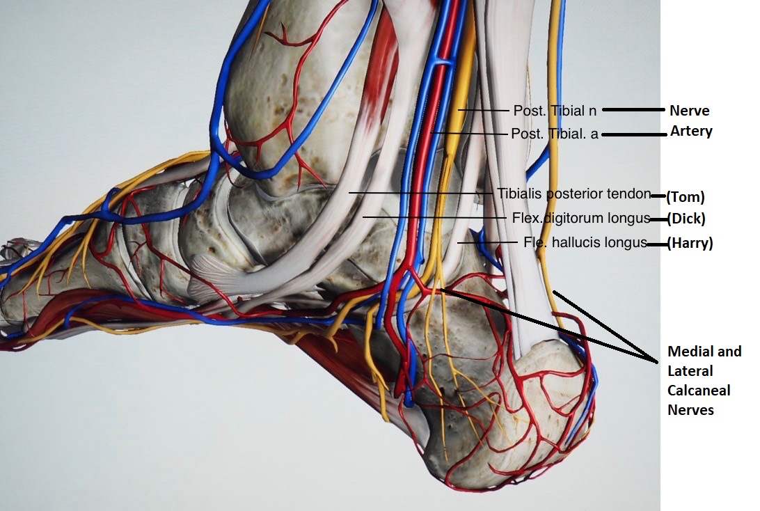
Photo credit: http://www.bats.ac.nz/detail-foot_and_ankle_surgery-2 with some labelling by Hannah Salsbury
The Other stuff:
In the picture above, the red lines are the blood vessels and dressed in blue, the veins! The arteries bring the blood downhill from the heart and the veins, they pump the blood back uphill to the lungs to start the cycle all over again. Any of these can get clogged by build-up around the edges or blocked by a rogue clot floating around your body.
Let’s not forget the nerves of the foot. The medial and lateral calcaneal nerves are pictured above in yellow. Nerves are where the brain sends sensory signals as well as the signal to fire the muscles. Think of them like electricity wires. They are verrry sensitive and can be injured easily.
There are fluid-filled sacks called bursae around the back of the heel that if they get inflamed can take up space and press on the nerves giving us pain in the heel. Anything taking up more space than it is meant to be bound to cause trouble.
I hope this helps people understand that the foot may be more complicated than they anticipated. All these structures work so beautifully together over our whole lifetime….except when they are NOT working. We can’t just jump to conclusions about what might be causing your heel pain. So how the hell do you find out what is going on and more importantly- “When will I get better” you say. Well, you are going to have to read on…..
Next week- What is a Podiatrist going to do about my sore heel??
References:
1. Brukner, P & Khan, K 2007, Clinical Sports Medicine, 3rd edition, Tata McGraw Hill, Australia.
2. Cole, C, Seto, G & Gazewood, J 2005, ‘Plantar Fasciitis: Evidence-Based Review of Diagnosis and Therapy`, American Family Physician, vol. 72, no. 11, pp. 2237-42.
3. Thomas, JL, Christensen, JC, Kravitz, SR, Mendicino, RW, Schuberth, JM, Vanore, JV, Weil, LS, Zlotoff, HJ, Bouche, R & Baker, J 2010, ´ The Diagnosis and Treatment of Heel Pain: A Clinical Practice Guideline–Revision 2010`, The Journal of Foot & Ankle Surgery, vol. 49, pp. 1-19.
4. McMinn, M.H, Hutchings, R. T, Logan, B.M, Colour Atlas of Foot and Ankle Anatomy 2nd edition, Mosby-Wolfe, Spain.
5. L. Kelly, S, Kuitunen, Racinais, A, Cresswell. Recruitment of Plantar Intrinsic Foot Muscles with Increasing Postural Demand. Clinical Biomechanics 27 (2012) 46-51
Sorry, the comment form is closed at this time.